 NanoFCM


Quantification of Functionally Accessible Ligands on LNP Surfaces
Author: Meiwafosis Co., Ltd. (In collaboration with Nitto Denko Corporation)Overview
Expanding the therapeutic scope of mRNA requires delivery systems with precise cell selectivity. While ligand-functionalized lipid nanoparticles (LNPs) enable targeted delivery, the quantitative relationship between multivalent surface avidity and delivery efficiency has remained unresolved.
This study established a single-domain antibody (VHH)-functionalized LNP platform with controlled orientation and tunable ligand density. By combining a fluorescent antigen-binding assay with single-particle nanoflow cytometry, we selectively quantified functionally accessible ligands on the LNP surface.
This approach demonstrated that CD8-targeted LNPs exhibit a bell-shaped delivery efficiency profile, peaking at an optimal density of approximately 0.1 VHH per 100 nm². LNPs displaying the optimal functional VHH density achieved selective mRNA expression in CD8⁺ T cells, enabling in vivo CAR-T cell generation.
Design of VHH-Modified LNPs
Design of VHH Ligands
Full-length IgG antibodies are relatively large and contain multiple potential sites for conjugation to the LNP surface. As a result, their orientation on the LNP surface is difficult to control precisely. In contrast, genetically engineered single-domain antibodies (VHHs) can be designed to contain a single, site-specific conjugation handle, enabling controlled orientation on the LNP surface. Their small size also facilitates efficient removal of unconjugated VHH molecules.
Preparation of VHH-LNPs and Bulk Quantification of VHH Ligands
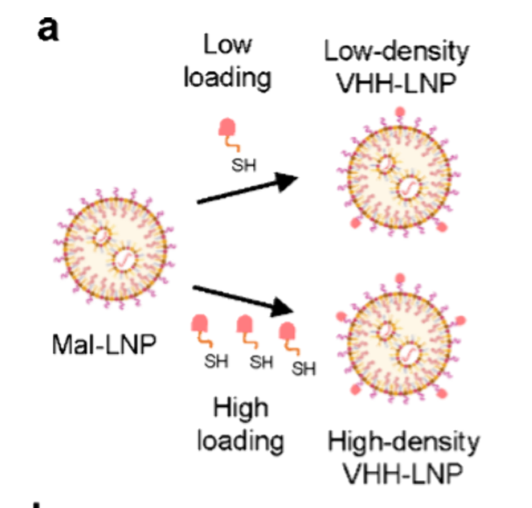
An anti-human CD8 VHH containing a cysteine residue was conjugated to maleimide-functionalized LNPs. By increasing the VHH input stepwise from 0.008 to 0.5 mol% relative to total lipid, VHH-LNP formulations with different ligand densities were prepared (Figure 1).
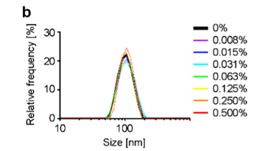
Physicochemical characterization demonstrated comparable hydrodynamic diameters (77-87 nm), low PDI values (<0.141), high mRNA encapsulation efficiencies (>96%), and similar apparent pKa values (6.08-6.15) across all formulations. These results indicate that tuning the surface ligand density did not compromise the physicochemical properties or overall quality of the LNP formulations (Figure 2).
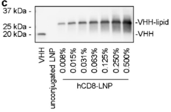
SDS-PAGE and Western blot analysis showed that the intensity of the VHH-conjugated lipid band increased with increasing VHH input (Figure 3). These results indicate that the amount of LNP-conjugated VHH increased with the VHH input level.
Total protein content in purified LNPs was also quantified using a BCA assay (Figure 4). Consistent with the Western blot results, the measured protein amount increased with the input VHH level.
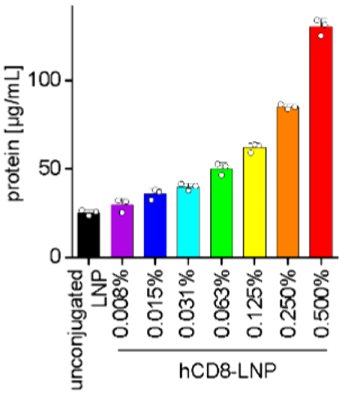
However, the conventional bulk assays shown in Figures 3 and 4 cannot distinguish functionally accessible ligands from total ligands, nor can they resolve particle-to-particle heterogeneity.
Quantification of Surface VHH Density on LNPs by NanoFCM
Nanoflow cytometry overcomes the size detection limits of conventional flow cytometry, enabling high-fidelity characterization of individual LNPs.
Anti-human CD8 VHH-LNPs (hCD8-LNPs) were incubated with an Alexa Fluor 488 (AF488)-labeled recombinant human CD8 protein probe. Functionally accessible VHH molecules on the LNP surface bind the fluorescently labeled recombinant CD8 probe, thereby enabling selective detection of functionally accessible ligands (Figure 5).
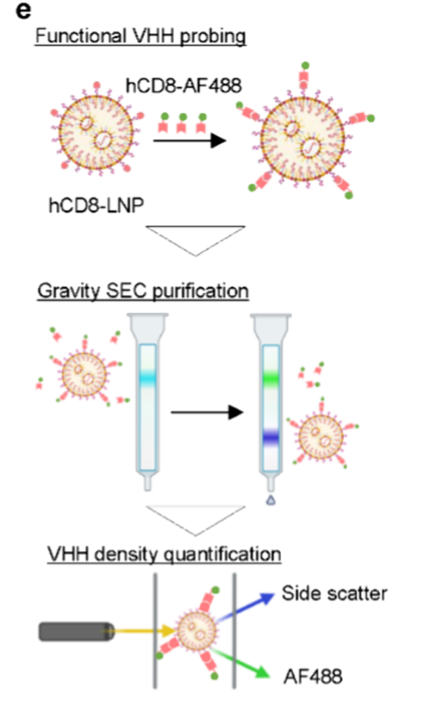
Individual LNPs were analyzed by NanoFCM to simultaneously measure AF488 fluorescence and side-scatter signals.
Calibration with size standard beads and molecules of equivalent soluble fluorochrome (MESF) calibration beads enabled conversion of side-scatter and fluorescence signals into particle diameter and the estimated number of bound VHH molecules, respectively. Each dot represents an individual LNP (Figure 6).
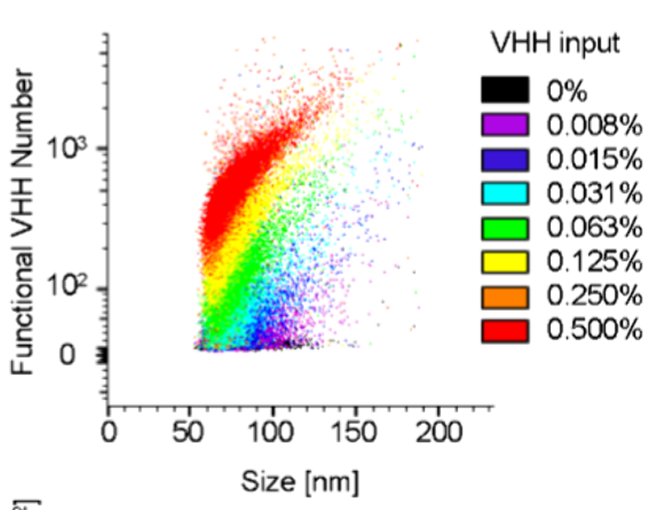
Because larger LNPs carry more VHH molecules, the number of bound VHH molecules was normalized to the surface area of each particle to calculate the VHH density per 100 nm² of particle surface (Figure 7).
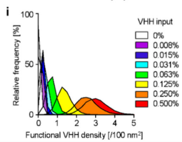
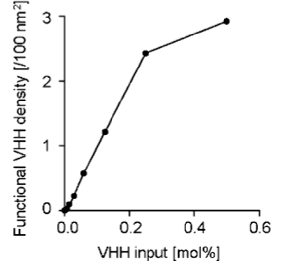
Functional ligand density increased proportionally with the input VHH amount up to 0.25 mol% (Figure 8). At 0.5 mol%, the surface ligand density reached a plateau, suggesting saturation of available conjugation sites or steric hindrance at the LNP surface.
Evaluation of Cellular Delivery Efficiency of VHH-LNPs
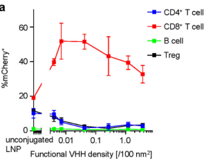
The effect of surface VHH density on the efficiency of targeted mRNA delivery to CD8⁺ T cells was evaluated. Human PBMC-engrafted mice were intravenously administered VHH-LNPs encapsulating mCherry mRNA. mCherry expression in splenic CD8⁺ T cells reached its maximum with LNPs displaying an intermediate VHH density (0.042 VHH/100 nm²). Delivery efficiency diminished at both lower and higher VHH densities, demonstrating a bell-shaped relationship between ligand density and in vivo delivery efficiency (Figure 9).
In Vivo Validation
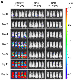
Anti-CD19 CAR mRNA was encapsulated in hCD8-LNPs formulated with the optimal ligand density, and the antitumor efficacy of in vivo-generated CAR-T cells was evaluated in a mouse model co-engrafted with Nalm6-GFP/Luc leukemia cells and human PBMCs. Two intravenous doses of CAR mRNA-loaded hCD8-LNPs were administered following cell engraftment. Control animals received hCD8-LNPs encapsulating mCherry mRNA.
Bioluminescence imaging demonstrated near-complete suppression of tumor growth in groups treated with CAR mRNA-loaded hCD8-LNPs, even at a dose as low as 0.1 mg/kg, whereas rapid tumor progression was observed in the control group (Figure 10). These findings demonstrate that in vivo-generated CAR-T cells achieve potent antigen-specific cytotoxicity and systemic therapeutic efficacy.
Conclusion
Collectively, these findings demonstrate that increasing ligand density enhances multivalent binding, whereas supra-optimal ligand densities limit functional mRNA delivery to target cells. The original study showed that excessive ligand density promotes CD8 receptor internalization and degradation, thereby reducing receptor availability for subsequent LNP binding and uptake.
This phenomenon was observed not only in LNPs formulated with the CL4F8-6 ionizable lipid but also in LNPs formulated with the clinically validated ALC-0315 ionizable lipid. The consistency of these findings across distinct ionizable lipid backbones and targeting formats supports the broad applicability of this mechanistic principle to antibody-functionalized LNP design.
This study establishes surface density as a tunable and quantifiable determinant of targeted mRNA delivery. The nanoscale ligand-density analysis developed in this study provides a quantitative framework for the mechanism-guided design of antibody-functionalized LNPs.
Reference:
HASHIBA, Kazuki, et al. Balancing Multivalent Avidity and Receptor Availability Governs mRNA Delivery by Antibody-Functionalized Lipid Nanoparticles. Nano Letters, 2026, 26.14: 4630-4641.
Link to the Original Research Article:
https://pubs.acs.org/doi/10.1021/acs.nanolett.5c06525
Related News from Nitto Denko Corporation:
