Notice: Undefined index: HTTP_ACCEPT_LANGUAGE in /www/wwwroot/www.nanofcm.com/wp-content/plugins/NanoFCMEditor/main.php on line 25
Liposomes – NanoFCM

Doxorubicin-carrying Liposomes

Author: admin     Date: February 22, 2024

Characterization of Doxorubicin-Carrying Doxoves

In nanomedicine development, nanoparticles are utilized as carriers to deliver payloads such as therapeutic agents. Therefore, the simultaneous detection of both nanoparticles and their cargos is desirable. Doxil (doxorubicin-carrying liposomes) is the first FDA-approved nanomedicine (1995), and DLS and cryo-TEM are the two most commonly used methods for size analysis. While DLS is not suitable for heterogeneous samples, the three-dimensional (3D) reconstruction of cryo-TEM usually takes 2-3 days.

1676874366895020.webp

Learn More(点击跳转)

Multiparameter Quantification of Liposomes

A liposome is a tiny vesicle made from materials similar to a cell membrane. Drug-encapsulated liposomes have been considered the most clinically acceptable drug delivery systems. In this study, the Flow NanoAnalyzer was used to simultaneously detect the side-scatter and fluorescence signals generated by individual liposome particles at a speed of up to 10,000 nanoparticles/min. To address the size dependence of the refractive index of liposomes, different sizes of doxorubicin-loaded liposomes were fabricated and characterized to serve as calibration standards for measuring both particle size and drug content. This method provides a highly practical platform for the characterization of liposome vesicles.

1676874477385924.webp

Learn More(点击跳转)


Characterization of Theranostic Liposomes

Author: admin     Date: February 22, 2024

Stimuli-responsive nanotheranostic systems, integrated with diagnostic and treatment features, have recently emerged and attracted much interest. However, most of the research focuses on the novelty of nanomaterials and undervalues the significance of single-particle characterization, which can provide detailed physical and biochemical information for performance evaluation and heterogeneity assessment. Due to the small particle size and low content of functional modules, high throughput and multiparameter analysis of individual stimuli-responsive nanoparticles remains challenging.

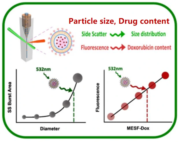
Here, using a reactive oxygen species (ROS)-responsive liposome (Lipo@BODIPY11) as an example, a strategy for characterizing theranostic nanoparticles with the Flow NanoAnalyzer was developed. Coincident detection of light scatter and fluorescence intensity provided a measurement for liposome quality assessment. Theranostic performance, referring to stimuli-responsive capability and drug release behavior upon ROS treatment, was obtained in minutes.

1678348773763716.webp

Figure 1. Flourescent analysis of Lipo@BODIPY11 by Flow NanoAnalyzer

1678348777370881.webp

Figure 2. Simultaneous detection of the ROS sensing and controlled drug release behavior of Lipo@BODIPY11&MXT by Flow NanoAnalyzer

This Flow NanoAnalyzer-based method provides a comprehensive approach for the proof-of-principle study, heterogeneity assessment, and quality control of biochemical nanosensors and theranostic nanomaterials.

Biosens. Bioelectron., 2019, 131, 185-192.

Quantification of Available Ligand Density on the Surface of Targeted Liposomal Nanomedicines

Author: admin     Date: February 22, 2024

Active targeting has been hailed as one of the most promising strategies to further enhance the therapeutic efficacy of liposomal nanomedicines. Owing to the critical role of ligand density in mediating cellular uptake and the intrinsic heterogeneity of liposomal formulations, precise quantification of the surface ligand density on a single-particle basis is of fundamental importance. In this work, we report a method to simultaneously measure the particle size and the number of ligands on the same liposomal nanoparticles by nanoflow cytometry. From this the ligand density for each individual liposome could be determined. The correlation between the available ligand density and cell targeting capability was assessed in quantitatively for liposomes modified with three different targeting moieties. The optimal ligand density was determined to be 0.5–2.0, 0.7, and 0.2 ligands per 100 nm^2 for folate-, transferrin-, and HER2-antibody-conjugated liposomes, respectively. These optimal values agreed well with the spike density of the natural counterparts, viruses. The as-developed approach can be applied to a wide range of active-targeting nanocarriers. The correlation between the available ligand density and cell targeting capability was assessed in a quantitative perspective for liposomes modified with three different targeting moieties.

1678349404355437.webp

1678349409810047.webp

                                                   Figure 1. Nano-flow cytometry (nFCM) analysis                               Figure 2. Measurement and calculation of the ligand 

                                                        of the surface folate receptor expression                                             density by nano-flow cytomstry (nFCM)

The optimal ligand density was determined to be 0.5–2.0, 0.7, and 0.2 ligands per 100 nm^2 for folate-, transferrin-, and HER2-antibody-conjugated liposomes, respectively. These optimal values agreed well with the spike density of the natural counterparts, viruses. The as-developed approach is generally applicable to a wide range of active-targeting nanocarriers.

ACS Nano, 2022, 16(4), 6886-6897.