Notice: Undefined index: HTTP_ACCEPT_LANGUAGE in /www/wwwroot/www.nanofcm.com/wp-content/plugins/NanoFCMEditor/main.php on line 25
Exosomes from various sources – NanoFCM

Analysis of Physicochemical Properties of EVs from Different Sources

Author: admin     Date: February 20, 2024

Physical Properties: Size Distribution and Particle Concentration

EV content has been gaining increasing interest from the EV community, primarily mRNAs, miRNAs and proteins. In addition, particular markers exposed on the lipid bilayers that determine specific interactions with target cells. However, it has been suggested that physical properties of  EVs may also affect their behavior, such as, the way they mediate intercellular communication.

Learn More(点击跳转)

Protein Analysis

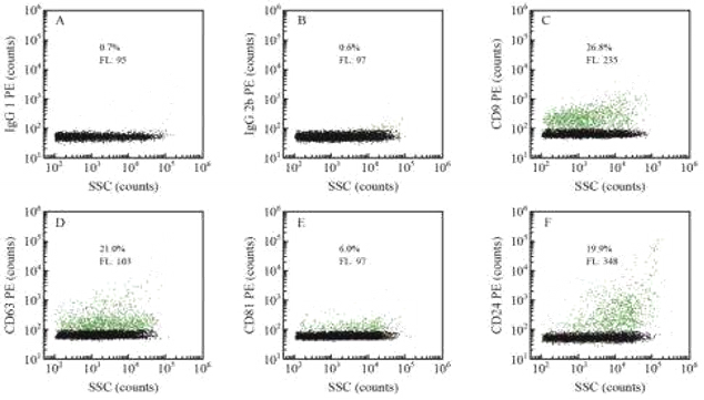
The biomolecular cargo (that is, proteins, nucleic acids and lipids) of EVs is thought to reflect their origin, suggesting that the cargo could be a promising source for the discovery of novel biomarkers. Although the EVs research has not matured to the point where it can propose a list of EV-specific “markers” that distinguish subsets of EVs from others, several tetraspanins, especially CD63, CD81, and CD96 have been used as markers of exosomes for the last two decades, due to their accumulation in small EVs as compared to whole cell lysates, and to the steady-state accumulation of CD63 in MVBs. As per guidelines from International Society for Extracellular Vesicles, ISEV, these markers can be used to differentiate  EVs from non-EVs.

Learn More(点击跳转)

Lipid and Nucleic Acids Analysis

Early studies suggested that EVs perform protein transport functions, specifically targeting receptor cells and exchanging proteins and/or lipids to trigger downstream signalling events.  It was discovered by researchers in 2007 that EVs are also capable of transporting nucleic acids and are involved in intercellular communication. EVs play a role in intercellular communication and organismal regulation through signalling molecules such as proteins and lipids on the membrane, as well as EV contents (neurotransmitters, enzymes, hormones and nucleic acids, etc.) encapsulated within the membrane. Therefore, in addition to studying the function of proteins, it is necessary to perform a comprehensive multi-parameter characterisation of EVs at single particle level.

Learn More(点击跳转)


Multi-parameter Characterization of Bacterial Vesicles

Author: admin     Date: February 20, 2024

Bacterial membrane vesicles (BMVs) are nano-scale lipid vesicles secreted by bacteria during the growth process. BMVs carry nucleic acids and proteins, especially virulence factors and immune regulatory factor for the inter and intra-species communication. BMVs not only affect a variety of biological processes, but have also shown important applications in vaccine and anti-cancer drug development. However, the purification and characterization of BMVs face serious challenges due to sample heterogeneity, small size and sparse contents. Obtaining a high-purity sample of BMVs and analysing this quantitatively poses another urgent challenge for BMVs research.

1676357291679198.webp

This study established a quantitative characterization method for the purity, particle concentration and size distribution of BMVs at the single particle level based on the Flow NanoAnalyzer. The separation and purification of BMVs was performed by combining differential centrifugation and density gradient centrifugation.

1677723152969683.webp

Figure 1. Purity and particle concentration of BMVs

1677723158397532.webp

Figure 2. Size distribution of BMVs.

In this study, a method was established to characterize the purity, particle concentration and size distribution of BMVs based on the Flow NanoAnalyzer. The Flow NanoAnalyzer provides a powerful platform for further research of biological function of BMVs.

CHINESE J ANAL CHEM, 2021, 49(5), 733-742.


Comprehensive Analysis of Urinary Extracellular Vesicles

Author: admin     Date: February 20, 2024

Urinary extracellular vesicles (uEVs) are used as a new class of liquid biopsy markers with promising applications in disease diagnosis and therapeutic monitoring. However, researchers lack knowledge about the particle size distribution, particle concentration, and biochemical phenotypes of uEVs at the nanoscale due to the lack of corresponding characterization techniques. EVs are highly individual,  implying an urgent need for the development of high-throughput, multi-parameter single particle detection technologies. In this study, multi-parameter quantitative characterization of uEVs purified by ultracentrifugation at the single particle level was performed using the Flow NanoAnalyzer to obtain comprehensive information on their purity, concentration, particle size, and contents. 

  1677723966109294.webp

Figure 1. The size distribution of uEVs

1677723972352927.webp


                                                             Figure 2. Purity and concentration analysis of uEVs                                Figure 3. Nucleic Acids analysis of uEVs                                               

image.png

Figure 4. Protein marker analysis of uEVs

In this study the positive rates of uEVs for CD9, CD63, CD81 and CD24 were 26.8%, 21.0%, 6.0% and 19. 9%, respectively. The Flow NanoAnalyzer may provides a platform to help further the research of the biochemical properties and clinical application of uEVs.

CHINESE J ANAL CHEM, 2021, 48(10), 1305-1314.


Multi-parameter characterization for tear fluid EVs

Author: admin     Date: February 20, 2024

Extracellular vesicles (EVs) in easily accessible body fluids have emerged as a promising source for liquid biopsy diagnosis. Although the collection of tears can be fast, safe, and noninvasive, the researches on EVs in tear fluid are rare. Additionally, the involvement of the EVs in physiological and pathological processes is still largely unknown. The aim of this study was to analyze and characterize EVs in tear fluid at single-particle level to reveal the population heterogeneity by the Flow NanoAnalyzer. The expression of CD9, CD63, CD81, CD47, CD45, CD24, and EpCAM was assessed via immunofluorescent detection. The EV concentration and size distribution in tear fluid was measured using SSC light detection. It also compared the positive rate and abundance of tissue factor (TF) in tear and saliva EVs, providing theoretical support for further exploration of the function of tear EVs.

1678168844215104.webp

Figure 1. Morphology and purity characterization of EVs isolated from unstimulated tears.

1678168869260977.webp

              Figure 2. Concentration analysis and particle size distribution of EVs in tears.               Figure 3. Protein marker phenotyping 

                                                                                                                                                     of EVs isolated by UC from human tears.

1678168894374074.webp

Figure.4. Characterization of TF-exposing EVs in tear and saliva.

The purity, size distribution, particle concentration and the expression of eight membrane proteins of tear fluid EVs were analysed using the Flow NanoAnalyzer. Moreover, the presence of high TF expression in tear EVs exhibited strong coagulation activity, providing a new research perspective for ophthalmic studies.

CHINESE J ANAL CHEM, 2021, 48(10), 1305-1314.