Notice: Undefined index: HTTP_ACCEPT_LANGUAGE in /www/wwwroot/www.nanofcm.com/wp-content/plugins/NanoFCMEditor/main.php on line 25
Fundamental Research – NanoFCM

Label-Free Detection of 27 nm MS2

Author: admin     Date: February 21, 2024

Viruses are by far the most abundant biological entities on our planet. While animal viruses are notoriously responsible for many fatal diseases, plant viral pathogens cause significant losses in crop yields worldwide each year. On the other hand, the high transfection efficiency of viral gene-therapy vectors and the monodisperse structures that have a precise shape and size make viral particles powerful drug delivery vehicles and versatile nanotechnological building blocks. Therefore, high-resolution and high-throughput analysis of single viral particles are in high demand for virology research, disease diagnosis and treatment, and biotechnology and nanotechnology applications.

However, due to the small size (mostly ranging from 20-200 nm in diameter) and simple structure of viruses, conventional flow cytometry, which focuses on particles larger than 200 nm, is not the best option for virus detection. There is a great necessity for the development of advanced approaches enabling rapid, label-free, and accurate detection of viruses at the single-particle level. The development of the Flow NanoAnalyzer opens a new avenue for virus characterization
1678342479868977.webp

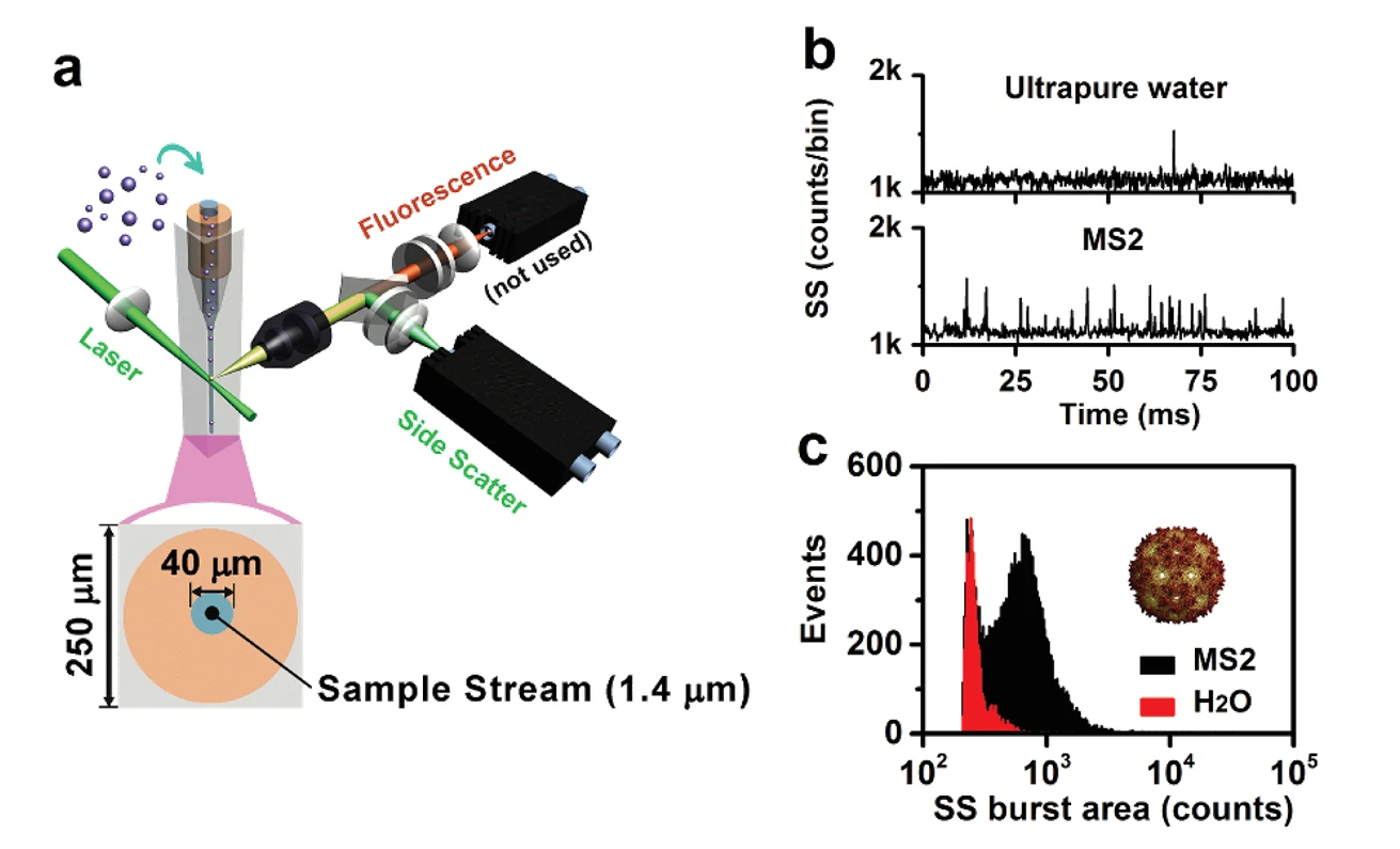
Figure 1. Label-free detection of levivirus MS2 by the Flow NanoAnalyzer.

The virus used in this experiment is levivirus MS2, which is a non-enveloped, spherical virion about 27 nm in diameter. The genome is monopartite, linear ssRNA (+), approximately 3.5 kb in size. The signal-to-noise (S/N) ratio, calculated as the average burst height of all the nanoparticles detected in 1 min divided by the standard deviation of the background signal (noise), is 11 for the MS2 viruses. This indicates that the Flow NanoAnalyzer provides exceptional sensitivity in discerning MS2 viruses against the background noise. This level of sensitivity could meet the detection demands of most virus nanoparticles in nature.

Angew. Chem. Int. Edit., 2016, 55(35), 10239-10243.

Particle size analysis

Author: admin     Date: February 21, 2024

Effect of the Refractive Index on the Light Scattering Detection

Based on the theory of Rayleigh scattering, the scattering cross-section of a nanoparticle is not only dominated by the particle size but also influenced by the refractive index contrast, which is the ratio of the refractive index of the particle to that of the medium. A silica nanoparticle scatters 3.8-fold less light than a polystyrene nanoparticle of the same size at a wavelength of 532 nm, with refractive indices of 1.46 and 1.59, respectively.

The exact refractive index of viruses is not known, but it can be assumed to be 1.45 or 1.46, similar to that of proteins and DNA. The effect of the refractive index on the light scattering detection of single viruses is examined using the bacteriophage T7, a bacterial virus with an icosahedral capsid head of ~60 nm in diameter, along with silica nanospheres and polystyrene nanobeads of a comparable size.

Learn More

Discrimination and Size Measurement of Viruses in a Mixture

Nano-sized biological agents and pathogens, such as viruses, are known to be responsible for a wide variety of human diseases, such as the flu, AIDS, and herpes. They have also been used as biowarfare agents. Viral size plays a substantial role in transmission dynamics, disease outbreaks, and outcomes. Rapid and accurate detection and characterization of single viral particles have become increasingly important for virology research, disease diagnosis and treatment, and biotechnology applications.

Learn More

Purity Assessment and Dynamic Monitoring of the Viral Genome Release Process

Author: admin     Date: February 22, 2024

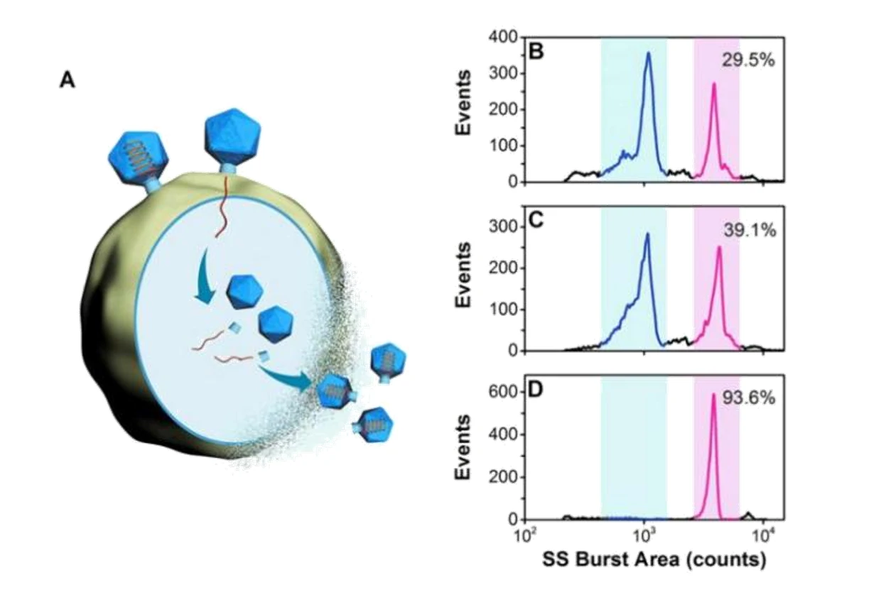
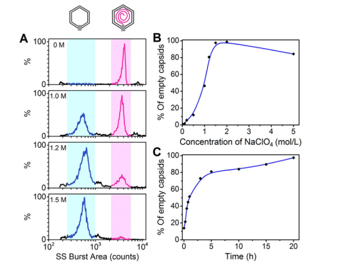
Quality control is indispensable to ensure the adequate purity of virus products in the biopharmaceutical industry. Purity assessment of virus products is crucial in many biotechnology applications. Many particles can coexist in the lysates of the host cells infected with a wild-type phage, including cell debris, DNA-free proheads, empty capsids (after DNA ejection), and mature virions. Meanwhile, the natural process of viruses delivering their genes into the hosts for self-replication has inspired the design of virus-like particles to deliver therapeutic or imaging agents. Monitoring the viral genome release process will allow for deeper insights into the mechanisms of cargo release. The Flow NanoAnalyzer is applied to analyze the composition of the viral sample and monitor the viral genome release process following treatment with NaClO4.

1678342874921878.webp

Figure 1. Analysis of crude samples for bacteriophage T7 from different batches.

1678342878270903.webp

Figure 2. Analysis of NaClO4-triggered DNA ejection process of bacteriophage T7

The differentiation of capsids before DNA packaging (proheads, ~1100 counts) and after DNA release (empty capsids, ~600 counts), indicates that the Flow NanoAnalyzer could be an efficient tool for virology study.

Angew. Chem. Int. Edit., 2016, 55(35), 10239-10243.