Notice: Undefined index: HTTP_ACCEPT_LANGUAGE in /www/wwwroot/www.nanofcm.com/wp-content/plugins/NanoFCMEditor/main.php on line 25
Bacteria Basic Research – NanoFCM

Autofluorescence Quantification of Single Bacteria

Author: admin     Date: February 22, 2024

Cellular autofluorescence in the visible region can affect the sensitivity of fluorescence microscopic or flow cytometric assays by interfering with or even precluding the detection of low-level specific fluorescence. On the other hand, detection of autofluorescence can provide information for bacterial discrimination and identification. The autofluorescence detected in the green region may orginate from flavins, which comprise a category of molecules that include riboflavin (RF, vitamin B2) and its derivatives flavin adenine dinucleotide (FAD) and flavin mononucleotide (FMN). The oxidized form of flavins all share remarkably similar spectral characteristics with fluorescein isothiocyanate (FITC). Three yellow-green fluorescent Fluospheres beads with different sizes and known fluorescein equivalents were analyzed in parallel with bacterial samples to construct the calibration curve between the mean fluorescence burst area and the FITC equivalents per nanoparticle. The burst area distribution histogram of bacterial autofluorescence was then converted to the distribution of FITC equivalents per bacterial cell.

image.png

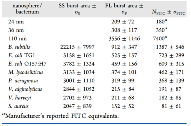
The Flow NanoAnalyzer can detect the autofluorescence of a single bacterium and demonstrate that the green autofluorescence mainly originates from the oxidized form of endogenous flavin.

Anal. Chem., 2012, 84(3), 1526-1532.

High-Throughput Single-Cell Analysis of Low Copy Number Protein

Author: admin     Date: February 22, 2024

Single-cell analysis is vital in providing insights into the heterogeneity of molecular content and phenotypic characteristics of complex or clonal cell populations. As many essential proteins and most transcription factors are produced at a low copy number, analytical tools with superior sensitivity to enable the analysis of low abundance proteins in single cells are in high demand. β-galactosidase (β-gal) has been the standard cellular reporter for gene expression in both prokaryotic and eukaryotic cells. Here, the Flow NanoAnalyzer is used for the development of a high-throughput method for the single-cell analysis of low copy number β-gal proteins. Upon fluorescence staining with a fluorogenic substrate C12FDG, quantitative measurements of the basal and near-basal expression of β-gal in single bacteria are demonstrated. Combined with the quantitative fluorometric assay and the rapid bacterial enumeration, the β-gal expression distribution profile could be converted from arbitrary fluorescence units to protein copy numbers per cell.

image.png

Built upon the sensitivity and speed of the instrument and the good cell retention of the hydrolysis, products of C12FDG, β-gal are detected at single bacterial cell level.

Biosens. Bioelectron., 2013, 48, 49-55.

Detection of protein interaction in bacterial two-hybrid system

Author: admin     Date: February 22, 2024

Characterization of protein-protein interactions (PPIs) is critical to understanding cellular signal transduction pathways. However, quantitative measurement of binding strength remains challenging. Based on the classic bacterial adenylate cyclase two-hybrid (BACTH) system, previous studies have confirmed that the relative reporter protein expression (RRPE) is related to the interaction between the two proteins and the binding strength-related intrinsic features.

In this study, the gene of the fluorescent protein tdTomato was inserted into the chromosome through CRISPR/Cas9 technology, and one of the interacting proteins was marked with a 12 amino acid tetracysteine (TC), which could be further marked with a transmembrane double arsenic dye. The combined use of TdTomato and the TC-tag enables rapid and high-throughput analysis of the expression levels of reporter proteins and protein interactions at the single-particle level by the Flow NanoAnalyzer, thus simplifying the quantitative measurement of PPI. In addition, the study also used the developed RRPE-tdTomato-TC-BACTH method, demonstrating that the method is sensitive enough to distinguish small differences with an affinity of 1.4 times and can quickly screen out protein interaction inhibitors.

1678353133996330.webp

Figure 1. Simultaneous detection of tdTomato–TC-BACTH protein expression

1678353137426178.webp

Figure 2. tdTomato-BACTH method for rapid identification of target cells

Combined with dual fluorescent labeling assays, the Flow NanoAnalyzer can eliminate fixation and permeabilization treatments, greatly speeding up sample analysis. Quantitative analysis of protein interactions and reporter protein expression was performed simultaneously. Based on the tdTomato-BACTH method of NanoFCM, the traditional agar plating step is eliminated. Thereby, providing high-throughput identification of target cells and verifying that the tdTomato-BACTH method has the potential of high-throughput PPI research.

Talanta, 2021, 233, 122549.