VIRUSES/VIRAL VACCINE
INTEGRATE INTO THE GENOMES OF THE HOST CELLS
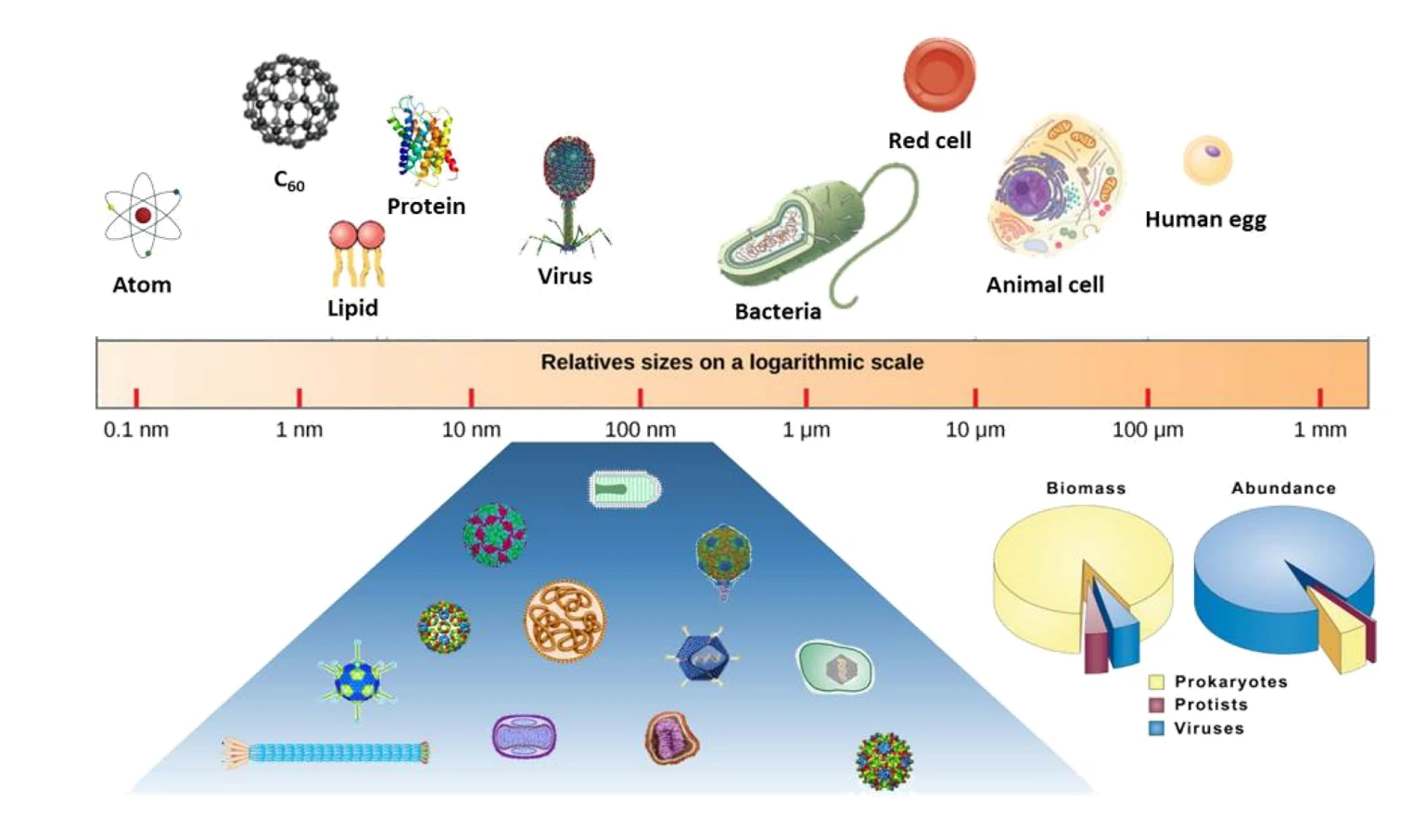
Virus is a general term for a group of tiny organisms that are made up of non-cellular forms of nucleic acid molecules (DNA or RNA) and proteins and live in parasitic life. Viruses are by far the most abundant ‘lifeforms’ in nature and are the reservoir of most of the genetic diversity in the oceans, and it is also the chief culprit of many fatal diseases. On the other hand, the extensive study of viruses as pathogens has yielded detailed knowledge about their biological, genetic and physical properties. Viruses bear exceptional stability and biocompatibility, they have attracted much attention due to the advantages of nanometer-scale size (20-200 nm), high degree of symmetry and polyvalence, and the relative ease of producing large quantities.

Moreover, the particles present programmable units, which can be modified by either genetic modification or chemical bioconjugation methods. Recently viruses are acting more positively, oncolytic viruses, a typical branch of new therapeutic agents, rely on both the selective tumor cell killing and the induction of systematic antitumor immunity to achieve antitumor responses, has shown great promise in tumor treatment. Chimeric antigen receptor (CAR) T cell therapy for B cell malignancies have unexpectedly fueled an increasing number of clinical trials and the US Food and Drug Administration’s first approval of cell therapies for cancer treatment. Moreover, adeno associated viruses (AAV) are common vectors for gene transfer in vivo and vectors with potential in therapeutic applications. Encouraging results of AAV vectors have been observed in preclinical models. To be specific, the delivery of CRISPR/Cas9 nucleases via AAV vectors displays therapeutic utility in preclinical models of a variety of diseases, and this approach is being rapidly applied to clinical trials. Rapid detection and accurate characterization of viruses has become increasingly important over the years. The physical and chemical properties of viruses, such as size, concentration, biochemical component as well as the packaging efficiency of functional molecules would cause direct effect to their application. Transmission electron microscopy (TEM) is still the most recognized means to measure the particle size and morphology of a virus despite the fact that it is labor intensive and time-consuming. Conventional plaque titer and TCID50 method are the most classical approaches for concentration measurement of viruses, however, they quantify only those which cause visible cell-damage thus exclude the viruses without infectivity. At present there does not exist any virus analysis method available to biologist which can quickly and reliably detect, quantify and characterize virus particles with single particle sensitivity. Thus it is highly desirable for the development of rapid and sensitive technique for the analysis of single vial nanoparticles. Among the many characterization methods, light scattering technology stands out as a sensitive, non-destructive single particle characterization method. The analysis of viral particles by flow cytometry based on light scattering has become the mainstream method for virus assays. In addition to early applications in counting environmental viruses, flow cytometry provides information on viral particle size, integrity and aggregation, genetics, lipid and protein composition, protein conformation, and so on. However, it is difficult for traditional flow cytometry to meet the needs of individual virus particle detection due to the limitation of sensitivity. Here, the Flow NanoAnalyzer is introduced, which can directly detect MS2 virus with particle size as low as 27 nm by light scattering. It provides an effective means for single particle multi-parameter characterization of viral particles.


