Notice: Undefined index: HTTP_ACCEPT_LANGUAGE in /www/wwwroot/www.nanofcm.com/wp-content/plugins/NanoFCMEditor/main.php on line 25
Nanometer materials – NanoFCM

Gold Nanoparticles

Author: admin     Date: February 23, 2024

Gold nanoparticles (AuNPs) have been a key focus in medical, bioanalytical, and catalytic applications due to their unique photo-thermal properties. Accurate particle size and concentration analysis are critical for quality control during AuNP synthesis, surface functionalization modification, and development analysis. Transmission electron microscopy (TEM) is the most commonly used method for morphological characterization of AuNPs particles. Dynamic light scattering (DLS) can quickly measure the particle size of AuNPs but can only measure uniform samples. However, compared with many methods, accurate and effective measurement of concentration is lacking. The concentration of AuNPs particles is typically analyzed by combining TEM and inductively coupled plasma mass spectrometry or spectroscopy (ICP-MS or ICP-AES). This method is not only time-consuming, and labor-intensive, but also leads to the deviation of the volume calculation due to the irregular shape and uneven particle size of the AuNPs, resulting in significant differences between the experimental and actual results. By means of single-particle counting and sample volume flow per unit time measurement, the absolute quantitative analysis method of particle concentration without standards was developed on the Flow NanoAnalyzer. In addition, via the multi-parameter detection performance of the instrument, a simple fluorescence internal standard quantitative method was developed.
1678429628211333.webp

Figure 1. Analysis of a single gold nanoparticle by the Flow NanoAnalyzer

The method for determining the concentration of AuNPs based on single particle counting is suitable for the detection of nanoparticles of various single or mixed materials. It solves problems related to the accurate concentration measurement of irregularly shaped, composite and hybrid nanoparticles.

J. Am. Chem. Soc., 2010, 132(35), 12176-12178.

Ag@sio2 Core-Shell Nanoparticles

Author: admin     Date: February 23, 2024

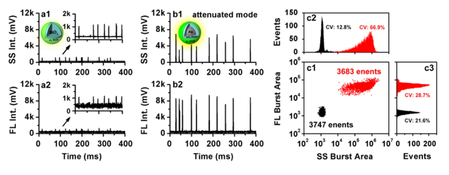
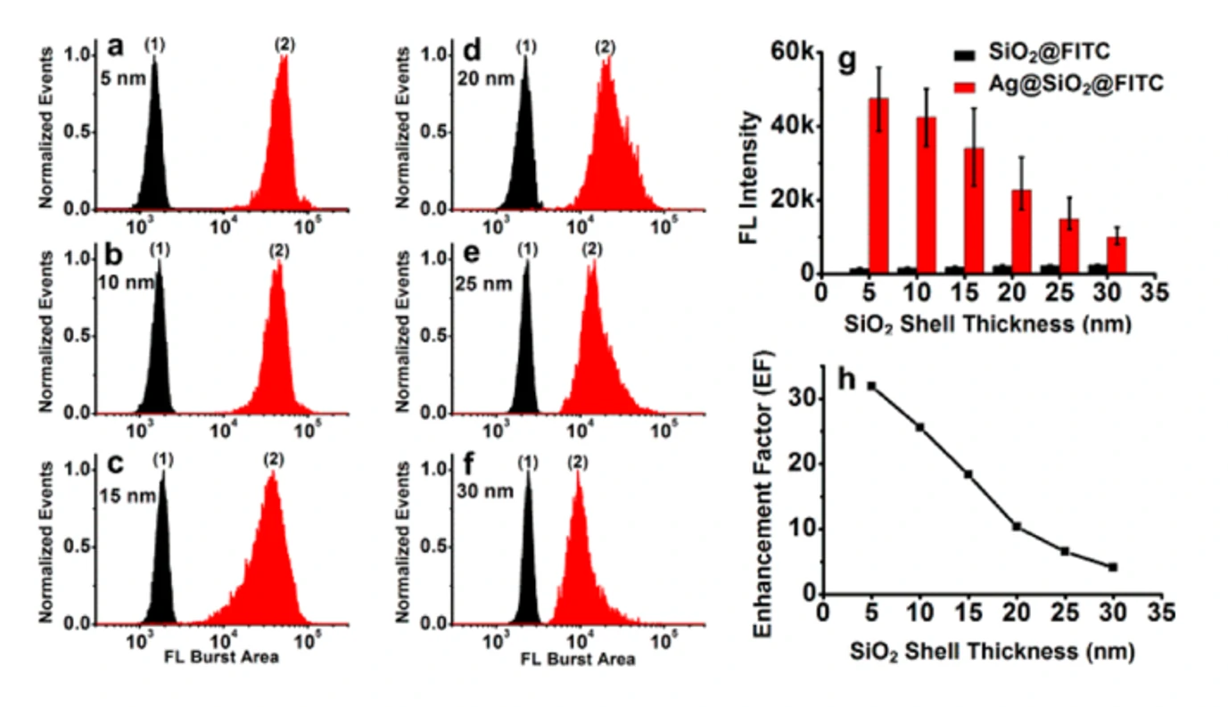
Metal-enhanced fluorescence (MEF) based on localized surface plasmon resonance (LSPR) is an effective method to improve the sensitivity of detection. Plasmonic nanoparticles are inherently heterogeneous, so single-particle analysis of MEF in free solution is key to understanding and controlling the MEF process. In this study, the Flow NanoAnalyzer was used to study the fluorescence enhancement of a single plasmonic nanoparticle near to fluorophore molecules. The study used Ag@SiO2 core-shell nanoparticles as a model system, which consist of a silver core, a silica shell, and a thin layer of FITC-doped silica shell. FITC-doped silica nanoparticles of the same size but with no silver nucleus as their counterparts were used. The Flow NanoAnalyzer was employed to detect side-scattering and fluorescence signals of single particles in the suspension and systematically study the effects of the size of silver nuclei (40-100 nm) and the distance between fluorophore and metal (5-30 nm).

1678429846391181.webp

Figure 1. Analysis of MEFs at the Single Particle Level by the Flow NanoAnalyzer

1678429849536374.webp

Figure 2. Analysis of Ag@SiO2 core-shell nanoparticles with a silver core size of 70 nm and different silica shell thicknesses by the Flow NanoAnalyzer

The experimental observations in this study at the single-particle level are well supported by finite-difference time-domain (FDTD) calculations compared with ensemble-averaged fluorescence spectral measurements. This is very important for the design and control of plasmonic nanostructures, which can effectively enhance the fluorescence.

ACS Sens., 2017, 2(9), 1369-1376.

Rapid and Quantitative Measurement of Single Quantum Dots

Author: admin     Date: February 23, 2024

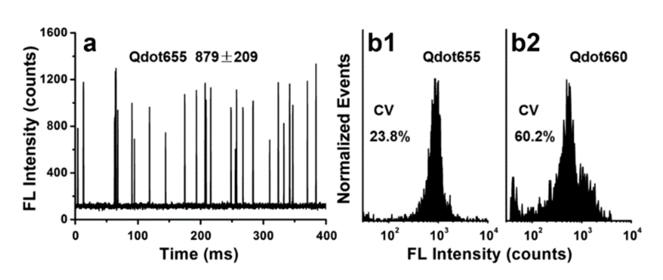
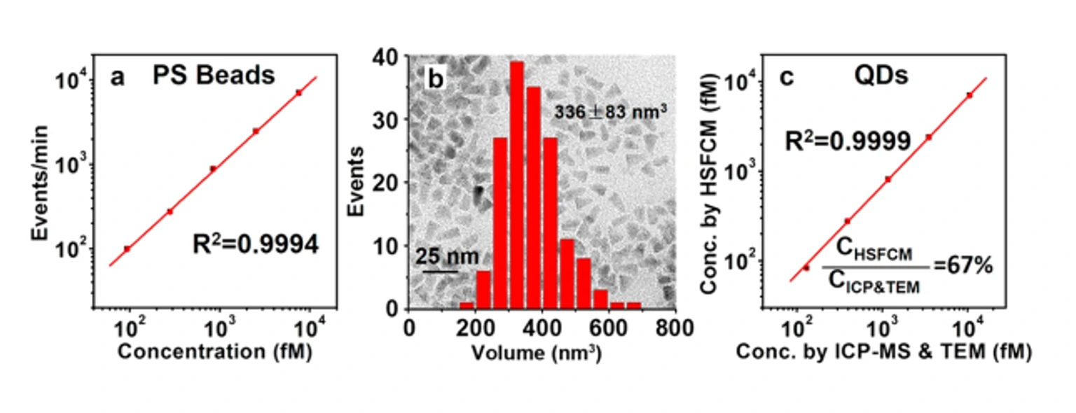
Semiconductor quantum dots (QDs) have a wide range of potential application in the biomedical field due to their strong fluorescence brightness and long-term photostability. This study reported a method for accurate quantification of the fluorescence intensity of single QDs using nano-flow cytometry. By analyzing thousands of QDs in less than one minute, this method quickly reveals the inherent polydispersity of QDs. The study also introduces the application of the Flow NanoAnalyzer in quality assessment of QDs, metal ions impact research, and coupling aggregation of biomolecular assessment. Through the single particle counting method, it enables the accurate measurement of the particle concentration of QDs.

1678430799748198.webp

Figure 1. Fluorescence quantitative analysis of a single QD

1678430802369339.webp

Figure 2. Particle concentration analysis of QDs

1678430806338030.webp

Figure 3. Statistical histograms of representative fluorescence burst trajectories and burst heights of Qdot655 in different buffers

Utilizing the Flow NanoAnalyzer, the fluorescence of individual QDs was rapidly quantified. Accurate quantification of QD concentration was achieved by single particle counting. Nano-flow detection provides a powerful characterization tool for the synthesis and application development of QDs.

Anal. Chem., 2017, 89(18), 9857-9863.

Light-Scattering Sizing of Single Submicron Particles

Author: admin     Date: February 23, 2024

The rapid detection of the particle size of a single submicron particle (100-1000 nm) is critical for the quality control of particulate matter, biomedical research, environmental research, and the development of drug delivery systems. Although the direct detection of elastically scattered light of a single submicron particle is the simplest way to measure particle size, the low sensitivity of detection instruments and the complex relationship between particle scattered light intensity and particle size make it a great challenge.

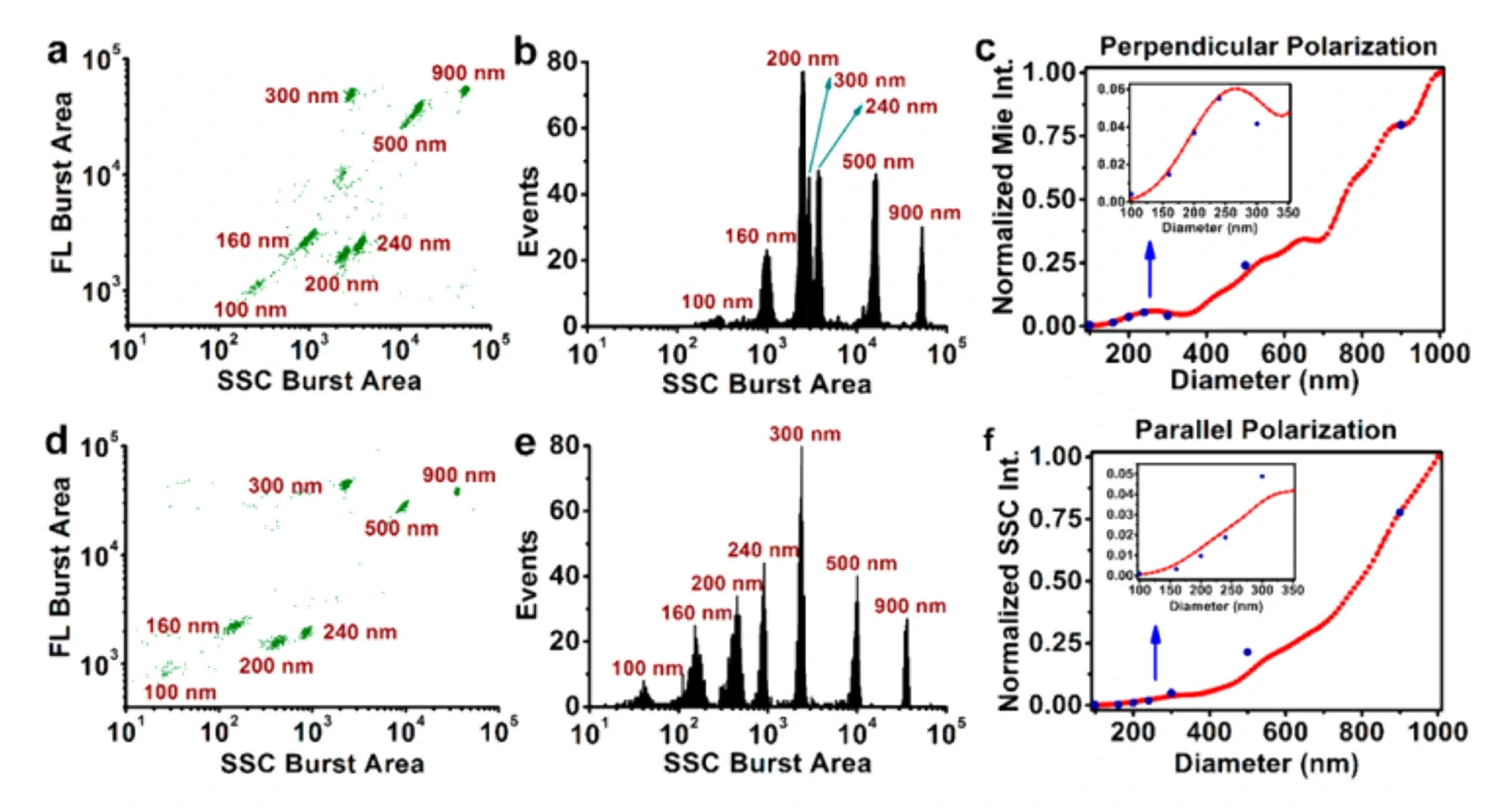
In this study, the high-level sensitivity of the Flow NanoAnalyzer is combined with the side scattering (SSC) detection of single nanoparticles, providing the first comparison of experimentally measured versus Mie theory calculated SSC light intensity of single submicron particles. Good consistency was observed between silica microspheres and polystyrene microspheres in both vertical and parallel polarization directions of the incident laser beam. Compared with vertical polarization, parallel polarization could be better resolved the microspheres of different sizes because the scattering intensity increases with the increase of particle size.

The prediction ability of the simple numerical model established in this work can be used to predict the scattering behavior of the Flow NanoAnalyzer. More importantly, using the linear relationship between the measured and the calculated scattering intensity, the researchers developed a method capable of measuring submicron particle sizes with the precision and accuracy of Mie theory, rather than a calibration curve fitted by several sparsely separated particle size reference standards. Therefore, this method has great potential in guiding the accurate measurement of submicron particle size.

1678431148179554.webp

Figure 1. Nano-flow cytometry detectors measure with perpendicular or parallel polarization of the laser. 

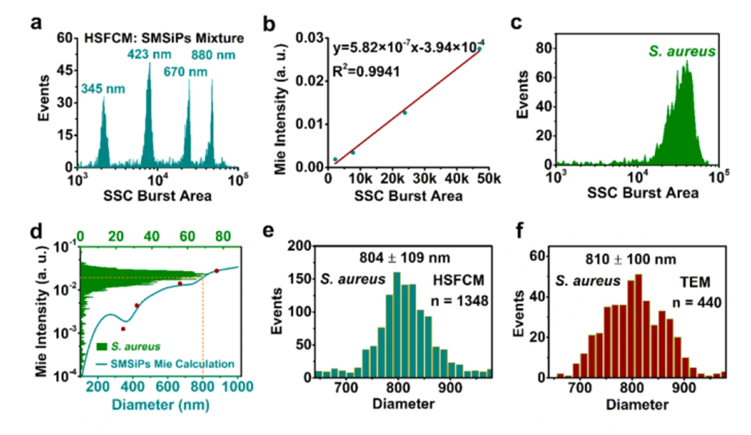
Mixture of commercially available fluorescent polystyrene microspheres from 100-900 nm

1678431152742711.webp

Figure 2. A new method for size measurement of Staphylococcus aureus 

based on nano-flow cytometry and Mie scattering calculations

Using the linear relationship between a particle's scattering intensity measured by the Flow NanoAnalyzer detector and the calculated scattering intensity, a method capable of measuring submicron particle size with the precision and accuracy of Mie theory was developed.

Anal. Chem., 2018, 90, 12768-12775.