
Flow NanoAnalyzer for Single Extracellular Vesicle Analysis
NanoFCM provides a versatile and powerful platform
Author: Mika Mika Date: December 2, 2021
The Flow NanoAnalyzer platform enables quantitative and multiparameter analysis of single extracellular vesicles. Which can detect the size distribution, concentration, biochemical properties and single Extracellular Vesicle Analysis.
Extracellular vesicles (EVs) are lipid bilayer-delimited particles that are naturally released from almost all types of cell and, unlike a cell, cannot replicate. They carry a cargo of proteins, nucleic acids, lipids, metabolites, and even organelles from the parent cell.
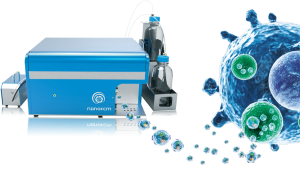
NanoFCM provides a versatile and powerful platform — Flow NanoAnalyzer. The platform is became a powerful tool for life science, nanoscience and nanotechnology studies. Flow NanoAnalyzer can be used for the multiparameter characterization of natural and synthetic nanoparticles (7-1000 nm) at the single-particle level. Combining light scattering and fluorescence detection, high-resolution distributions of particle size and biochemical properties can be acquired simultaneously in 1-2 minutes.
Flow NanoAnalyzer is the paltform for the multiparameter analysis of individual EVs. The fluorescence of single phycoerythrin molecules can be well detected against the background. The Flow NanoAnalyzer platform enables quantitative and multiparameter analysis of single EVs. The EVs size can be down to 40 nm, which is distinctively sensitive, yet high-throughput, and shows great potential in liquid biopsy applications.
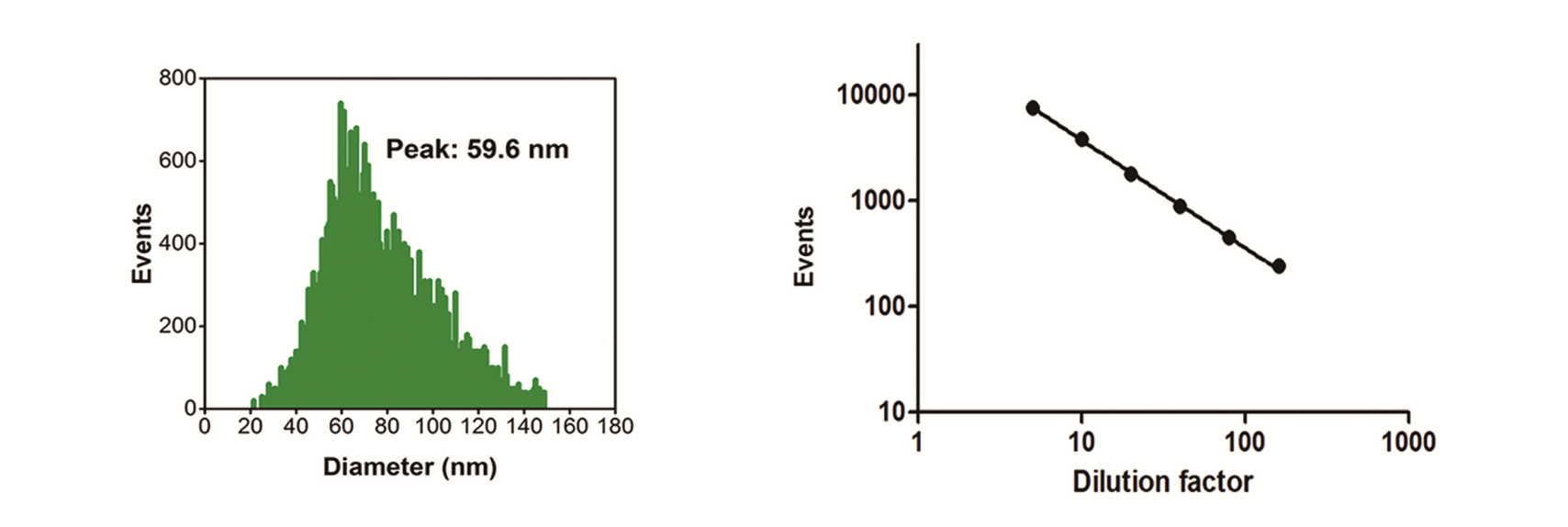
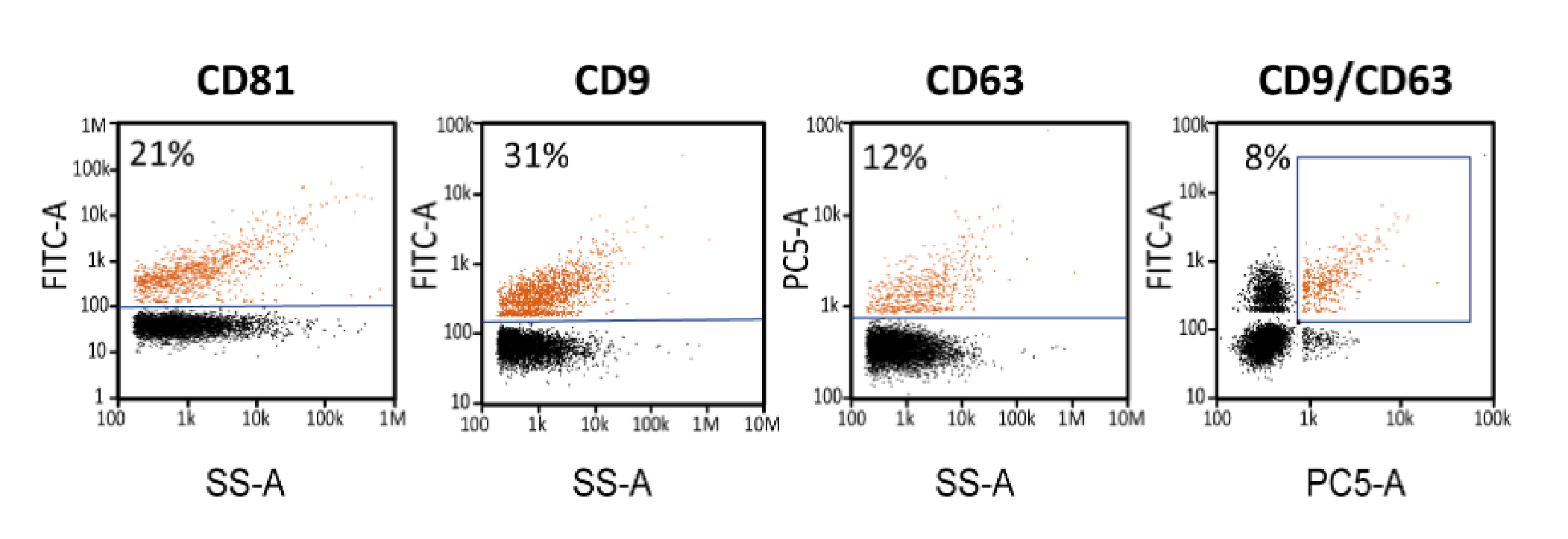
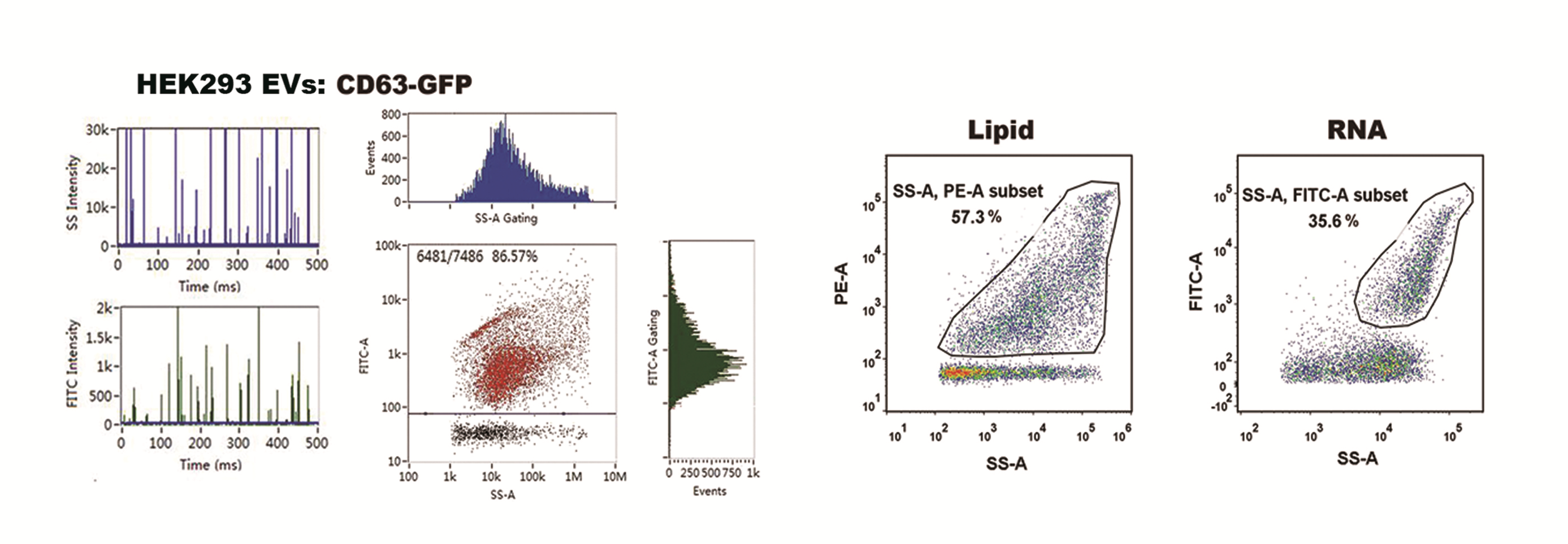
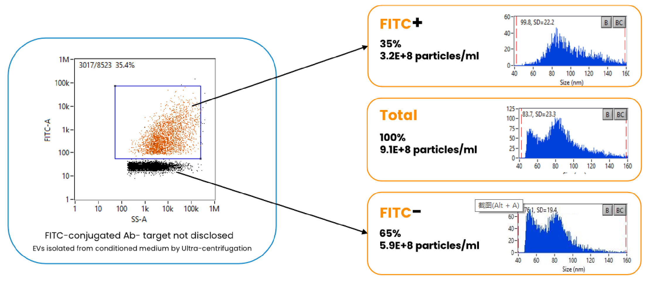
We have demonstrated the sensitivity of Flow NanoAnalyzer by detecting single silica nanoparticles. Silica nanoparticles have the similar refractive index with EVs. The fluorescence sensitivity of a single R-PE molecule has also been verified. The size (diameter) distribution and particle concentration of EVs could be acquired directly from the software. Expression of CD9, CD63, CD81 tetraspanins could be evaluated on the single extracellular vesicle level. Both intrinsic and extrinsic fluorescence could be detected individually. Colocalization of nucleic acids, surface markers and lipids can be achieved by simply dual-labeling. Monoclonal antibodies, aptamers and Annexin V are employed to label the surface markers and intraluminal molecules.